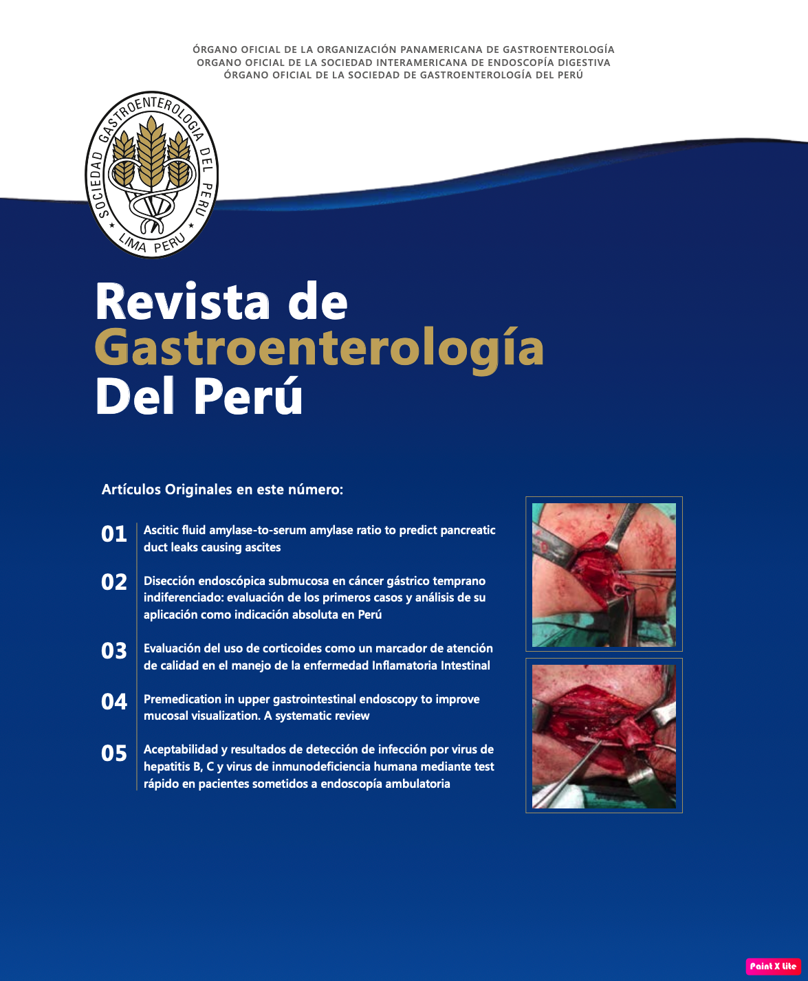
Complicaciones infrecuentes en un solo paciente: perforación esofágica, lesión vascular cervical e infección de tejidos blandos causadas por una espina de pescado
DOI:
https://doi.org/10.47892/rgp.2024.444.1753Palabras clave:
Migración de Cuerpo Extraño, Cuerpos Extraños, Esófago, Tracto Gastrointestinal Superior, Tomografía, Urgencias MédicasResumen
En este artículo, presentamos un caso clínico excepcionalmente raro y desafiante. Se trata de una mujer de 65 años que, mientras comía, ingirió accidentalmente una espina. Este cuerpo extraño, tras ser ingerido, migró desde el esófago proximal, hasta penetrar en la vena yugular interna izquierda. Este fenómeno inusual presentó como síntoma principal, disfagia alta de curso agudo, acompañada de un hematoma en el hemicuello izquierdo. Este caso no solo destaca la gravedad potencial de la ingesta accidental de cuerpos extraños, sino también la posibilidad de migración a localización infrecuentes potencialmente graves que conlleva a retos diagnósticos y terapéuticos. La migración de cuerpos extraños a través de los tejidos blandos y su posterior impacto en estructuras vasculares críticas son eventos extremadamente raros y requieren una intervención médica inmediata y especializada.
Descargas
Métricas
Citas
2. 3. Kavitha AK, Pinto O, Moras K, Lasrado S. An unusual migrated foreign body. J Clin Diagn Res. 2010;4:2903-2906.
Divya G, Hameed AS, Ramachandran K, Vinayak KV. Extraluminal migration of foreign body: a report of two cases. Int J Head Neck Surg. 2013;4(2):98-101. doi: 10.5005/jp-journals-10001-1150.
Jayachandra S, Eslick GD. A systematic review of paediatric foreign body ingestion: Presentation, complications, and management. Int J Pediatr Otorhinolaryngol. 2013;77(3):311-7. doi: 10.1016/j.ijporl.2012.11.025.
Pelucchi S, Bianchini C, Ciorba A, Pastore A. Unusual foreign body in the upper cervical oesophagus: case report. Acta Otorhinolaryngol Ital. 2007;27(1):38-40.
Benmansour N, Ouattassi N, Benmlih A, Elalami MN. Vertebral artery dissection due to an esophageal foreign body migration: A case report. Pan Afr Med J. 2014;17:1-2. doi: 10.11604/pamj.2014.17.96.3443.
Salil Kumar K, Rajan P, Muraleedharan Nampoothiri P, Jalaludhin J. Penetrating oesophageal foreign body. Indian J Otolaryngol Head Neck Surg. 2003;55(3):194-5. doi: 10.1007/BF02991953.
Koh WJ, Lum SG, Al-Yahya SN, Shanmuganathan J. Extraluminal migration of ingested fish bone in the upper aerodigestive tract: A series of three cases with broad clinical spectrum of manifestations and outcomes. Int J Surg Case Rep. 2021;89:106606. doi: 10.1016/j.ijscr.2021.106606.
Grayson N, Shanti H, Patel AG. Liver abscess secondary to fishbone ingestion: Case report and review of the literature. J Surg Case Reports. 2022;2022(2):rjac026. doi: 10.1093/jscr/rjac026.
Sierra-Ruiz M, Sáenz-Copete JC, Enriquez-Marulanda A, Ordoñez CA. Extra luminal migration of ingested fish bone to the spleen as an unusual cause of splenic rupture: Case report and literature review. Int J Surg Case Rep. 2016;25:184-7. doi: 10.1016/j.ijscr.2016.06.028.
Fan T, Wang CQ, Song YJ, Wu WY, Wei YN, Li XT. Granulomatous Inflammation of Greater Omentum Caused by a Migrating Fishbone. J Coll Physicians Surg Pakistan. 2022;32(8):S124-6. doi: 10.29271/jcpsp.2022.Supp2.S124.
Mulita F, Kehagias D, Tchabashvili L, Iliopoulos F, Drakos N, Kehagias I. Laparoscopic removal of a fishbone migrating from the gastrointestinal tract to the pancreas. Clin Case Reports. 2021;9(3):1833-4. doi: 10.1002/ccr3.3822.
Lee T-H, Park S-W, Ryu S, Cho KJ, Won SJ, Park JJ. Two cases of extraluminal migration of fishbones into the thyroid gland and submandibular gland. Ear, Nose Throat J. 2022;14556132210987. doi: 10.1177/01455613221098787.
Goh YH, Tan NG. Penetrating oesophageal foreign bodies in the thyroid gland. J Laryngol Otol. 1999;113(8):769-71. doi: 10.1017/s0022215100145165.
Chee LWJ, Sethi DS. Diagnostic and therapeutic approach to migrating foreign bodies. Ann Otol Rhinol Laryngol. 1999;108(2 I):177-80. doi: 10.1177/000348949910800213.
Remsen K, Biller HF, Lawson W, Som ML. Unusual presentations of penetrating foreign bodies of the upper aerodigestive tract. Ann Otol Rhinol Laryngol. 1983;92(4 Suppl. 105):32-44. doi: 10.1177/00034894830920s403.
Sekar R, Raja K, Ganesan S, Alexander A, Saxena SK. Migrated Foreign Body of Upper Digestive Tract—A Ten-Year Institutional Experience. Indian J Otolaryngol Head Neck Surg. 2022;74:5577-83. doi: 10.1007/s12070-021-02890-5.
Vadhera R, Gulati SP, Garg A, Goyal R, Ghai A. Extraluminal hypopharyngeal foreign body. Indian J Otolaryngol Head Neck Surg. 2009;61(1):76-8. doi: 10.1007/s12070-009-0039-z.
Zohra T, Ikram M, Iqbal M, Akhtar S, Abbas SA. Migrating foreign body in the thyroid gland, an unusual case. J Ayub Med Coll Abbottabad. 2006;18(3):65-6.
Osinubi OA, Osiname AI, Pal A, Lonsdale RJ, Butcher C. Foreign body in the throat migrating through the common carotid artery. J Laryngol Otol. 1996;110(8):793-5. doi: 10.1017/s0022215100134991.
Lue AJ, Fang WD, Manolidis S. Use of plain radiography and computed tomography to identify fish bone foreign bodies. Otolaryngol - Head Neck Surg. 2000;123(4):435-8. doi: 10.1067/mhn.2000.99663.
Drakos P, Ford BC, Labropoulos N. A systematic review on internal jugular vein thrombosis and pulmonary embolism. J. Vasc. Surg. Venous Lymphat. Disord. 2020;8(4):662-666. doi: 10.1016/j.jvsv.2020.03.003.
Agha R.A., Franchi T., Sohrabi C., Mathew G., Kerwan A., SCARE Group The SCARE 2020 guideline: updating consensus Surgical CAse REport (SCARE) guidelines. Int. J. Surg. 2020;84:226-230. doi: 10.1016/j.ijsu.2020.10.034.
Lin D, Reeck JB, Murr AH. Internal jugular vein thrombosis and deep neck infection from intravenous drug use: management strategy. Laryngoscope. 2004;114(1):56-60. doi: 10.1097/00005537-200401000-00009.
Kim JE, Ryoo SM, Kim YJ. Incidence and clinical features of esophageal perforation caused by ingested foreign body. Korean J. Gastroenterol. 2015;66(5):255-260. doi: 10.4166/kjg.2015.66.5.255.
ASGE Standards of Practice Committee; Ikenberry SO, Jue TL, Anderson MA, Appalaneni V, Banerjee S, et al. Management of ingested foreign bodies and food impactions. Gastrointest. Endosc. 2011;73(6):1085-1091. doi: 10.1016/j.gie.2010.11.010.
Ambe P, Weber SA, Schauer M. Swallowed foreign bodies in adults. Dtsch. Arztebl. Int. 2012;109(50):869-875. doi: 10.3238/arztebl.2012.0869.
Shrime MG, Johnson PE, Stewart MG. Cost-effective diagnosis of ingested foreign bodies. Laryngoscope. 2007;117(5):785-93. doi: 10.1097/MLG.0b013e31803c568f.
D’Costa H, Bailey F, McGavigan B, George G, Todd B. Perforation of the oesophagus and aorta after eating fish: An unusual cause of chest pain. Emerg Med J. 2003;20(4):385-6. doi: 10.1136/emj.20.4.385.
Brinster CJ, Singhal S, Lee L, Marshall MB, Kaiser LR, Kucharczuk JC. Evolving options in the management of esophageal perforation. Ann of Thorac Surg. 2004;77(4):1475-83. doi: 10.1016/j.athoracsur.2003.08.037.
Joshi AA, Bradoo RA. A foreign body in the pharynx migrating through the internal jugular vein. Am J Otolaryng. 2003;24(2):89-91. doi: 10.1053/ajot.2003.20.
Peng A, Li Y, Xiao Z, Wu W. Study of clinical treatment of esophageal foreign body-induced esophageal perforation with lethal complications. Eur Arch Oto-Rhino-L. 2012;269(9):2027-36. doi: 10.1007/s00405-012-1988-5.
Sengupta S, Kalkonde Y, Khot R. Idiopathic bilateral external jugular vein thrombosis–a case report. Angiology. 2001;52(1):69-71. doi: 10.1177/000331970105200110.
Chowdhury K, Bloom J, Black MJ. Spontaneous and nonspontaneous internal jugular vein thrombosis. Head Neck. 1990;12(2):168-173. doi: 10.1002/hed.2880120214.
Lee SH, Park JW, Han M. Internal jugular vein thrombosis with OHSS. J. Clin. Ultrasound. 2017;45(7):450-452. doi: 10.1002/jcu.22423.
Descargas
Publicado
Cómo citar
Número
Sección
Licencia
Derechos de autor 2025 Hernando Marulanda Fernández, Juan Sebastián Frías Ordoñez, Zoraida Contreras, Jorge Peñafiel Ruiz, William Otero Regino

Esta obra está bajo una licencia internacional Creative Commons Atribución 4.0.
Revista de Gastroenterología del Perú by Sociedad Peruana de Gastroenterología del Perú is licensed under a Licencia Creative Commons Atribución 4.0 Internacional..
Aquellos autores/as que tengan publicaciones con esta revista, aceptan los términos siguientes:
- Los autores/as conservarán sus derechos de autor y garantizarán a la revista el derecho de primera publicación de su obra, el cuál estará simultáneamente sujeto a la Licencia de reconocimiento de Creative Commons que permite a terceros compartir la obra siempre que se indique su autor y su primera publicación esta revista.
- Los autores/as podrán adoptar otros acuerdos de licencia no exclusiva de distribución de la versión de la obra publicada (p. ej.: depositarla en un archivo telemático institucional o publicarla en un volumen monográfico) siempre que se indique la publicación inicial en esta revista.
- Se permite y recomienda a los autores/as difundir su obra a través de Internet (p. ej.: en archivos telemáticos institucionales o en su página web) antes y durante el proceso de envío, lo cual puede producir intercambios interesantes y aumentar las citas de la obra publicada. (Véase El efecto del acceso abierto).


 2022
2022 