
Prevalence of Barrett’s esophagus and factors associated with the diagnosis of dysplasia or adenocarcinoma in patients evaluated at a Chilean university endoscopy center
DOI:
https://doi.org/10.47892/rgp.2025.454.2104Keywords:
Barrett Esophagus, Endoscopy, Gastrointestinal, AdenocarcinomaAbstract
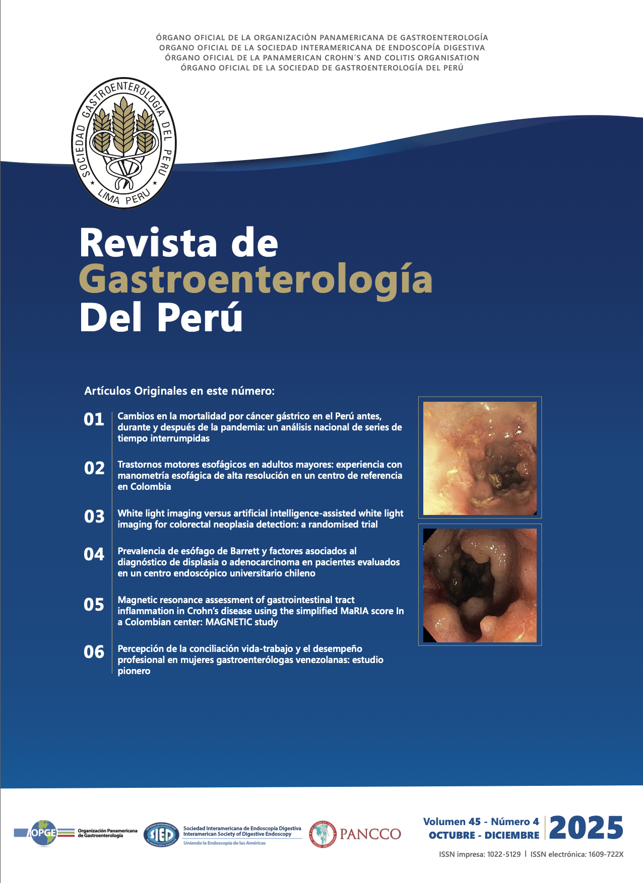
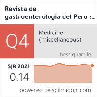
Introduction: Esophageal adenocarcinoma (EAC) is increasing in Western countries, and Barrett’s esophagus (BE) represents its only known premalignant condition. BE affects approximately 1–2% of the general population and up to 14% of patients with gastroesophageal reflux disease (GERD). Data from Latin America and Chile remain limited. Objectives: To determine the prevalence of BE, the neoplasia detection rate (NDR), and the endoscopic quality criteria associated with neoplasia detection in a Chilean university center. Materials and methods: A longitudinal cohort study including all patients with BE identified among upper gastrointestinal endoscopies performed at the Red de Salud UC CHRISTUS between January 2015 and December 2022. Patients with a history of other digestive neoplasms or referred with previously diagnosed BE/EAC were excluded. Demographic, endoscopic, and histopathological variables were analyzed. BE prevalence was defined as the number of histologically confirmed BE cases over the total diagnostic endoscopies performed during the study period. NDR was defined as the presence of highgrade dysplasia (HGD) or EAC on index endoscopy among BE patients. Multivariable logistic regression was applied to identify factors independently associated with NDR. Results: A total of 422 patients were diagnosed with BE (62% men; mean age 58 years, range 17-87). The overall prevalence of BE was 0.46% (422/91,723), increasing from 0.33% in 2015 to 0.72% in 2022. The low-grade dysplasia detection rate was 3.8% (16/422) and the NDR 1.7% (7/422). The mean BE length was 3,7 cm (range 1–18 cm). The Prague classification and chromoendoscopy were reported in 66% (280/422) and 44% (185/422) of procedures, respectively. Factors independently associated with neoplasia detection were age (OR 1.08; 95% CI 1.01-1.16), use of chromoendoscopy (OR 10.1; 95% CI 1.03-96), and presence of a visible lesion (OR 43.7; 95% CI 4.9-393). Conclusion: The prevalence of BE in this Chilean cohort was 0.46%, showing an upward trend approaching international reports. The use of chromoendoscopy and the detection of visible lesions were independently associated with higher neoplasia detection, underscoring the importance of adherence to endoscopic quality standards in BE evaluation.
Downloads
Metrics
References
Shaheen NJ, Falk GW, Iyer PG, Souza RF, Yadlapati RH, Sauer BG, et al. Diagnosis and management of Barrett’s esophagus: an updated ACG guideline. Am J Gastroenterol. 2022;117(4):559- 587. doi: 10.14309/ajg.0000000000001680
Weusten BLAM, Bisschops R, Dinis-Ribeiro M, di Pietro M, Pech O, Spaander MCW, et al. Diagnosis and management of Barrett esophagus: European Society of Gastrointestinal Endoscopy (ESGE) guideline. Endoscopy. 2023;55(12):1124-1146. doi: 10.1055/a-2176-2440.
Runge TM, Abrams JA, Shaheen NJ. Epidemiology of Barrett’s esophagus and esophageal adenocarcinoma. Gastroenterol Clin North Am. 2015;44(2):203-231. doi: 10.1016/j. gtc.2015.02.001.
Zagari RM, Eusebi LH, Rabitti S, Cristoferi L, Vestito A, Pagano N, et al. Prevalence of upper gastrointestinal endoscopic findings in the community: a systematic review of studies in unselected samples of subjects. J Gastroenterol Hepatol. 2016;31(9):1527-1538. doi: 10.1111/jgh.13308.
Eusebi LH, Cirota GG, Zagari RM, Ford AC. Global prevalence of Barrett’s oesophagus and oesophageal cancer in individuals with gastro-oesophageal reflux: a systematic review and meta-analysis. Gut. 2021;70(3):456-463. doi: 10.1136/ gutjnl-2020-321365.
Schneider JL, Corley DA. A review of the epidemiology of Barrett’s oesophagus and oesophageal adenocarcinoma. Best Pract Res Clin Gastroenterol. 2015;29(1):29-39. doi: 10.1016/j. bpg.2014.11.008.
Thrift AP, El-Serag HB, Kanwal F. Global burden and epidemiology of Barrett oesophagus and oesophageal cancer. Nat Rev Gastroenterol Hepatol. 2021;18(2):122-132. doi: 10.1038/s41575-021-00419-3.
Honing J, Fitzgerald RC. Categorizing risks within Barrett’s esophagus to guide surveillance and interception: suggesting a new framework. Cancer Prev Res (Phila). 2023;16(6):313-320. doi: 10.1158/1940-6207.CAPR-22-0447.
Kamboj AK, Katzka DA, Iyer PG. Endoscopic Screening for Barrett’s Esophagus and Esophageal Adenocarcinoma: Rationale, Candidates, and Challenges. Gastrointest Endosc Clin N Am. 2021;31(1):27-41. doi: 10.1016/j.giec.2020.08.002.
Antonios K, Aintabi D, McNally P, Berinstein E, Dutta P, Sampson N, et al. Risk Factors for the Development of Barrett’s Esophagus and Esophageal Adenocarcinoma: A Systematic Review and Meta-Analysis. Cancer Rep (Hoboken). 2025;8(3):e70168. doi:10.1002/cnr2.70168.
Rubenstein JH, Sawas T, Wani S, Eluri S, Singh S, Chandar AK, et al. AGA clinical practice guideline on endoscopic eradication therapy of Barrett’s esophagus and related neoplasia. Gastroenterology. 2024;166(6):1020-1055. doi: 10.1053/j. gastro.2024.03.019.
Chen Y, Sun C, Wu Y, Chen X, Kailas S, Karadsheh Z, et al. Do proton pump inhibitors prevent Barrett’s esophagus progression to high-grade dysplasia and esophageal adenocarcinoma? An updated meta-analysis. J Cancer Res Clin Oncol. 2021;147(9):2681-2691. doi: 10.1007/s00432-021- 03544-3.
Csendes A, Smok G, Sagastume H, Rojas J. Estudio prospectivo endoscópico y biópsico de la prevalencia de metaplasia intestinal en la unión gastroesofágica en controles y en pacientes con reflujo gastroesofágico. Rev Med Chil. 1998;126(2):155-161.
Csendes A, Smok G, Quiroz J, Burdiles P, Rojas J, Castro C, et al. Clinical, endoscopic, and functional studies in 408 patients with Barrett’s esophagus compared to 174 cases of intestinal metaplasia of the cardia. Am J Gastroenterol. 2002;97(3):554- 560.
Saha B, Vantanasiri K, Mohan BP, Goyal R, Garg N, Gerberi D, et al. Prevalence of Barrett’s esophagus and esophageal adenocarcinoma with and without gastroesophageal reflux: a systematic review and meta-analysis. Clin Gastroenterol Hepatol. 2024;22(7):1381-1394.e7. doi: 10.1016/j. cgh.2023.10.006.
Morgan E, Soerjomataram I, Rumgay H, Coleman HG, Thrift AP, Vignat J, et al. The global landscape of esophageal squamous cell carcinoma and esophageal adenocarcinoma incidence and mortality in 2020 and projections to 2040: new estimates from GLOBOCAN 2020. Gastroenterology. 2022;163(3):649– 658.e2. doi: 10.1053/j.gastro.2022.05.054.
Sugano K, Spechler SJ, El-Omar EM, McColl KEL, Takubo K, Gotoda T, et al. Kyoto international consensus report on anatomy, pathophysiology and clinical significance of the gastro-oesophageal junction. Gut. 2022;71(8):1488-1514. doi: 10.1136/gutjnl-2022-327281.
Kusano C, Singh R, Lee YY, Soh YSA, Sharma P, Ho KY, et al. Global variations in diagnostic guidelines for Barrett’s esophagus. Dig Endosc. 2022;34(7):1320-1328. doi: 10.1111/ den.14342.
Beaufort I, Akkerman E, van Munster S, Weusten B. Effect of biopsy protocol adherence vs nonadherence on dysplasia detection rates in Barrett’s esophagus surveillance endoscopies: a systematic review and meta-analysis. Endosc Int Open. 2023;11(3):E221-E229. doi: 10.1055/a-1967-1589.
Kolb JM, Davis C, Williams JL, Holub JL, Shaheen N, Wani S. Wide variability in dysplasia detection rate and adherence to Seattle protocol and surveillance recommendations in Barrett’s esophagus: a population-based analysis using the GIQuIC national quality benchmarking registry. Am J Gastroenterol. 2023;118(5):900-904. doi: 10.14309/ajg.0000000000002102.
Zagari RM, Eusebi LH, Galloro G, Rabitti S, Neri M, Pasquale L, et al. Attending training courses on Barrett’s esophagus improves adherence to guidelines: a survey from the Italian Society of Digestive Endoscopy. Dig Dis Sci. 2021;66(9):2888- 2896. doi: 10.1007/s10620-020-06615-6.
Iwaya Y, Iijima K, Hikichi T, Amano Y, Endo M, Goda K, et al. Evaluating the discrepancies between evidence-based and community standard practices in the endoscopic examination of Barrett’s esophagus: a nationwide survey in Japan. Esophagus. 2025;22(3):349-359. doi: 10.1007/s10388-025- 01127-6.
Inoue M, Ragunath K. Quality indicators in Barrett’s endoscopy: Best is yet to come. Dig Endosc. 2024;36(3):265-273. doi: 10.1111/den.14654.
Subhaharan D, Kakkadasam R, Ramaswamy P, Jones M, John S. Implementing educational interventions and key performance measures sustains quality of endoscopic assessment in patients with Barrett’s esophagus. Endosc Int Open. 2025;13:a25420618. doi: 10.1055/a-2542-0618.
Emura F, Chandrasekar VT, Hassan C, Armstrong D, Messmann H, Arantes V, et al. Rio de Janeiro Global Consensus on Landmarks, Definitions, and Classifications in Barrett’s Esophagus: World Endoscopy Organization Delphi Study. Gastroenterology. 2022;163(1):84-96.e2. doi: 10.1053/j. gastro.2022.03.022.
Redston M, Noffsinger A, Kim A, Akarca FG, Rara M, Stapleton D, et al. Abnormal TP53 predicts risk of progression in patients with Barrett’s esophagus regardless of a diagnosis of dysplasia. Gastroenterology. 2022;162(2):468-481. doi: 10.1053/j. gastro.2021.10.038.
Menon S, Norman R, Iyer PG, Ragunath K. Stratification of Barrett’s esophagus surveillance based on p53 immunohistochemistry: a cost-effectiveness analysis by an international collaborative group. Endoscopy. 2024;56(10):727- 736. doi: 10.1055/a-2317-8184.
Dhaliwal L, Codipilly DC, Gandhi P, Johnson ML, Lansing R, Wang KK, et al. Neoplasia detection rate in Barrett’s esophagus and its impact on missed dysplasia: results from a large population-based database. Clin Gastroenterol Hepatol. 2021;19(5):922-929.e1. doi: 10.1016/j.cgh.2020.07.034.
Kolb JM, Davis C, Williams JL, Holub J, Shaheen N, Wani S. High Rates of Dysplasia in a Population-based Analysis of “Incidental” Barrett’s Esophagus. Clin Gastroenterol Hepatol. 2025:S1542-3565(25)00189-2. doi: 10.1016/j.cgh.2025.01.017.
Downloads
Published
How to Cite
Issue
Section
License
Copyright (c) 2025 Alberto Espino, María Jesús Fuenzalida, Gonzalo Latorre, Felipe Silva, Oscar Corsi, Renato Palma, Javiera Torres, Rodrigo Nieto, Valentina Constanzo, Gabriela Fuentes, Mario Nilo, Leonardo Salgado, Cristóbal Vargas, José Ignacio Vargas

This work is licensed under a Creative Commons Attribution 4.0 International License.

Revista de Gastroenterología del Perú by Sociedad Peruana de Gastroenterología del Perú is licensed under a Licencia Creative Commons Atribución 4.0 Internacional..
Aquellos autores/as que tengan publicaciones con esta revista, aceptan los términos siguientes:
- Los autores/as conservarán sus derechos de autor y garantizarán a la revista el derecho de primera publicación de su obra, el cuál estará simultáneamente sujeto a la Licencia de reconocimiento de Creative Commons que permite a terceros compartir la obra siempre que se indique su autor y su primera publicación esta revista.
- Los autores/as podrán adoptar otros acuerdos de licencia no exclusiva de distribución de la versión de la obra publicada (p. ej.: depositarla en un archivo telemático institucional o publicarla en un volumen monográfico) siempre que se indique la publicación inicial en esta revista.
- Se permite y recomienda a los autores/as difundir su obra a través de Internet (p. ej.: en archivos telemáticos institucionales o en su página web) antes y durante el proceso de envío, lo cual puede producir intercambios interesantes y aumentar las citas de la obra publicada. (Véase El efecto del acceso abierto).


 2022
2022 